Neuroscience has advanced significantly thanks to the latest technological advances in the field. Scientists are now able to use light to selectively activate or deactivate neurons, independent of type or location, using a technique called optogenetics. Although intricate to implement, optogenetics offers an unprecedented degree of freedom to selectively manipulate specific parts of the brain while entirely avoiding others. This technique has enabled researchers to look at the brain less invasively and could offer valuable insight on its functions and the mechanisms behind mental health conditions such as anxiety disorders.
This year’s annual Beatty Memorial Lecture was held on October 16 at the Centre Mont-Royal, and featured Karl Deisseroth, a professor of bioengineering, psychiatry, and behavioural sciences at Stanford, as the speaker. Aside from his clinical work as a psychiatrist, he is an active researcher and a pioneer of optogenetics. This technique first took form in his lab just over a decade ago, and ever since, Deisseroth has continuously sought to improve neuroscience research by engineering better optogenetic methods and addressing classic questions in neuropsychiatry in new ways.
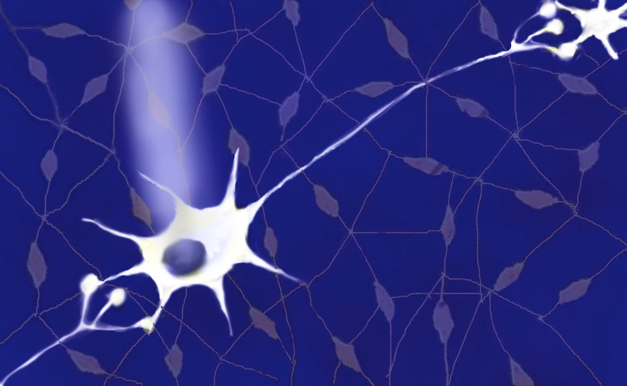
Optogenetics is a combination of genetics and optics to control the behaviour of certain cells. In neuroscience, optogenetics is used to control neurons by turning them on and off by exposing them to a certain colour of light. The opsins are inserted into the neurons using either microinjections or transgenic techniques. These opsins are light-sensitive pores, also referred to as light-sensitive ion channels, that form on the membrane and are sensitive to varying wavelengths of light. When the affected neurons are exposed to light, the pores either open, allowing ions to cross the membrane (exciting the neuron), or close, stopping ions from passing (inhibiting the neuron). This mimics the natural behaviour of these neurons and the way they connect with other neurons in the brain.
As a psychiatrist and a clinician, Deisseroth understands the importance of studying mental disease. During the lecture, he told the audience that “psychiatric disorders are not only chronic and fatal, but also still poorly understood.” Through his clinical work, he has learned that though there are distinct patterns in psychiatric disease that can be studied through patients’ subjective reports, measuring biological changes is a much more difficult task.
Is it possible then for optogenetics to help us understand neurological and psychiatric problems? The answer is yes. By exclusively activating a particular group of cells and thus the brain circuit they belong to, we can look at how they influence changes in behaviour patterns. By mapping these circuits and seeing what happens when they’re on and off, we can get closer to understanding how brain activity becomes affected by certain pathological conditions.
“Psychiatric disorders are not only chronic and fatal, but also still poorly understood.” – Karl Deisseroth, professor of bioengineering, psychiatry, and behavioural sciences at Stanford.
Deisseroth provided some examples of how this technique can give us insight into disorders like depression, Parkinson’s, and anxiety, by opening and closing circuits that researchers believe may be impaired. Previous research pointed toward a pattern of neural activity that may be altered in anxiety disorders. In a study published in Neuron, researchers were able to reduce anxiety in mice using optogenetics. They were able to insert the opsins into a specific subset of neurons, and when light was shone directly onto the brain to activate these opsins, animal tests showed reduced anxiety. Once the light was turned off, the animal went back to normal. These results show us how we can activate very specific cells and circuits in an animal’s brain and study what happens when they are turned on and off.
Deisseroth also went on to describe a second visualization technique called CLARITY that can make the brain transparent by taking away its fats, which block light from passing. Its novelty comes from the fact it does not require the brain or a sample block of tissue to be sliced into several thin slices to be imaged, and because antibody labels can also be viewed in the images. Additionally, the three-dimensional images produced are more detailed. However, this imaging technique requires either brain tissue to be extracted or has to be conducted post-mortem.
Neurons activated by mere flashes of light, entire brains made perfectly transparent — one can only be amazed at the innovative ideas springing up this millennium. In fact, the technique of optogenetics fits perfectly with the name of the talk: Illuminating the Brain, we illuminate the brain with different wavelengths of lights, but we also shine light on what may be actually happening inside of it. These techniques and the work of these researchers is definitely bringing us one step closer to understanding what’s happening inside the brain of the people that suffer poorly understood diseases. The hope is that this will one day allow us to create new treatments that target the specific circuits responsible for the brain abnormalities, instead of using chemical cocktails that affect the entire brain.